Transmission Muon Microscope by muon microbeam, realizing 3-D Imaging
[Purpose and Background of the Research]
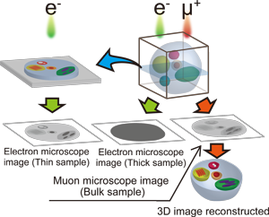
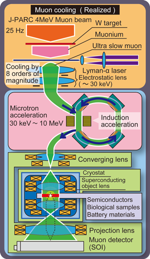
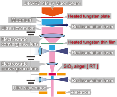
When a surface muon (4 MeV energy) impinges on a tungsten (or silica aerogel) foil, muonium (bound state of a positive muon with an electron, which is like a light isotope of hydrogen) is formed at the surface and evaporates into vacuum with a yield of around 4% (7%). By cooling muons by 7-8 orders of magnitude from 4 MeV to 0.2 eV (0.03 eV) combined with the application of the laser resonant ionization method (1p-2p-unbound) to efficiently ionize muonium atoms, ultra slow muon (USM) can be generated. Then re-accelerating and focusing USM help to achieve muon as a coherent wave. The essence of this research is to create a high intensity muon microbeam with excellent time and spatial resolution by using supercooling and re-acceleration to demonstrate the wave-particle duality of the muon. It will also provide a novel insight (3D imaging) to materials research (see Fig. 1).
 |  |
|---|---|
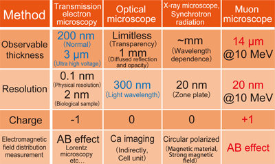
Fig. 1. 3D imaging of a 10 μm thick sample | Fig. 2. Comparison of Surface Analysis Methods |
USM will be re-accelerated up to 300 keV by induction acceleration to demonstrates the wave nature of the muon, and finally up to 10 MeV to get muon microbeam.
Using pulsed muon microbeam obtained by the re-acceleration of ultra-low energy muons, we will directly demonstrate the quantum coherence of the muon for the first time. In order to establish a transmission muon microscope, the following four research steps are required:
(A) Muon re-acceleration: Muons are re-accelerated up to 300 keV by induction acceleration, to demonstrate the wave nature of the muon. Furthermore, muon microbeam will be realized by further acceleration up to 10 MeV by microtron accelerator.
(B) Superconducting objective lens: Development of a superconducting objective lens that converges muons that are 200 times heavier than electrons.
(C) Transmission imaging: Development of normal conducting convergence projection lenses and muon image detector.
(D) Sample preparation: Preparation and observation of samples by making use of the high sample permeability of the transmission muon microscope.
 |
|---|
Fig. 3. Transmission muon microscope configuration diagram |
[Expected Research Achievements and Scientific Significance]
(1) Direct proof of quantum coherency of second generation lepton:
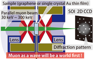
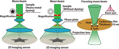
To demonstrate the muon as wave, a sample made of a single-crystal gold thin film will be used to observe a muon diffraction image. A gold grating (408 pm) with diffraction angles of 0.66 and 0.38 mrad for the acceleration voltages of 100 and 300 keV, respectively, will show diffraction patterns at a distance of 1 m on a two-dimensional image sensor SOI detector (Silicon on Insulator, 14 μm resolution) compatible with vacuum. It will be a historical achievement to be able to prove the quantum coherency exhibited by the muon as a second generation lepton in the standard model.
 |  |
|---|---|
Fig. 4. | Fig. 5. |
Fig. 4. Diffraction experiment by re-accelerated muon
Fig. 5. Muon microscopic imaging with re-accelerated muon
(2) Neuron network:
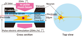
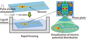
The network of neurons extending to several tens of micrometers and the three-dimensional structure of synapses with scale beyond that can be visualized with ns to μs time resolution by using an environmental cell and monitoring the change in the internal potential distribution of the electrical pulse response of a living nerve cell. For one-shot phenomena, we can also visualize the internal potential distribution of cells frozen rapidly at several tens of thousands of degrees per second.
 |  |
|---|---|
Fig. 6. | Fig. 7. |
Fig. 6. Visualization of neuron under electrical stimulation synchronized with accelerator
Fig. 7. 3D observation of rapidly frozen sample immediately after electrical stimulation
(3) Semiconductor device materials:
The muon phase difference imaging method has a shorter wavelength than that of an electron beam, and can obtain a larger phase variation. Especially, visualizing the internal potential distribution of a device with good reproducibility is important, making it possible to measure the internal potential distribution of semiconductor quantum dots in a solar cell with a thickness of several μm. It is also expected to be a diagnostic method to explore aspects that hinder the extraction of excited carriers by taking advantage of pulsed light irradiation synchronized with the accelerator.
(4) Crystal grain boundaries of batteries and magnetic materials:
By observing crystal grain boundaries (~10 μm in thickness) of microcrystal aggregates such as batteries and magnets, the dynamics of grain boundaries can be directly observed when an electromagnetic field is applied. In the field of batteries, the shape change of the electrode material accompanying charge and discharge, the formation of a secondary phase at the electrode/electrolyte solution interface, etc., affect the efficiency, life and reliability of the battery, and an in-situ observation technique is required. The high sample permeability of the muon enables observation of a Li ion battery in its environmental cell filled with electrolyte during normal electrode operation.
[Development of muon multi-step cooling method]
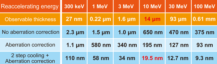
Spatial coherence (emittance) is improved by multi-step cooling, and imaging with nm-scale resolution can be realized. By repeating the muon cooling process by laser dissociation, and muon re-acceleration, reconvergence and re-impingement, the beam can be focussed to nm diameter. In the muon multi-step cooling method, the emittance is improved by two orders of magnitude or more in a single-step cooling process, and in a three-step cooling, the resolution could reach approximately 1 nm. The demonstration of the principle of the transmission muon microscope has the highest priority, and then three-dimensional observation is planned with atomic resolution up to the depth of 10 μm.
 |
|---|
Fig. 8. Transmission imaging ability of the transmission muon microscope |
 |
|---|
Fig. 9. Muon multi-step cooling method |